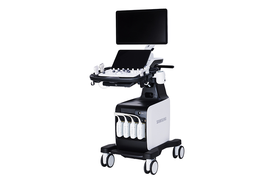
Embracing efficiency in your daily ultrasound scanning
Begin your journey towards efficient healthcare with the Samsung V6 ultrasound system. Our robust solution for general imaging offers both image clarity and advanced automated features. Additionally, Samsung’s cutting-edge imaging engine, Crystal Architecture™ ensures a reliable ultrasound experience.

 |
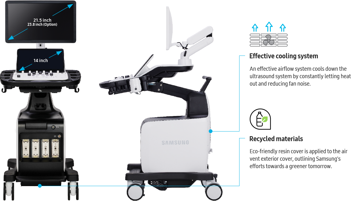
Experience simplicity with our easy-to-use system, specifically designed to alleviate your workload and enhance usability. Furthermore, our powerful system comes with battery capability, providing additional operational convenience. The Samsung V6 ultrasound system is a partner you can depend on to deliver exceptional efficiency to meet your daily ultrasound needs. |
![]()
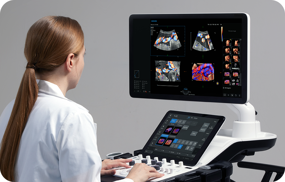
Elevating confidence with
superb imaging performance
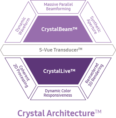 |
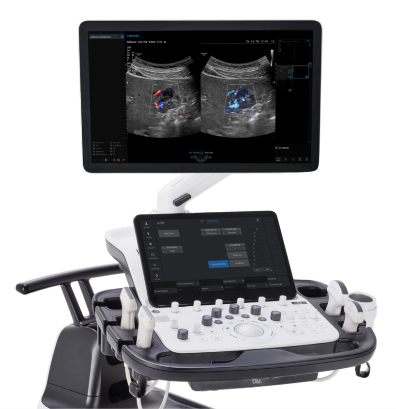
The V6 delivers exceptional 2D and color image quality tailored for general imaging, driven by Samsung’s core imaging engine, Crystal Architecture™. With its comprehensive imaging capabilities, the V6 is designed to seamlessly support your daily ultrasound scanning needs, enabling clear and accurate image acquisition. Experience confidence and accuracy in ultrasound scanning with the V6. |
Features
Reduce noise to improve 2D image quality
ClearVision enhances the edge contrast and creates sharp 2D images for optimal diagnostic performance.
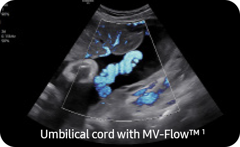
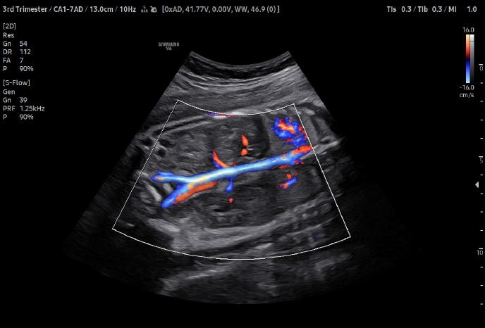
Visualize slow flow in microvascular vessels
MV-Flow™ visualizes microcirculatory and slow blood flow to display the intensity of blood flow in color.
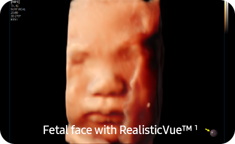
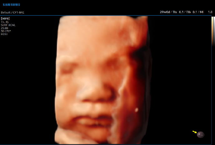
Express 3D anatomy with detail and realism
RealisticVue™ ¹ displays high resolution 3D anatomy with detailed expression and realistic depth perception.
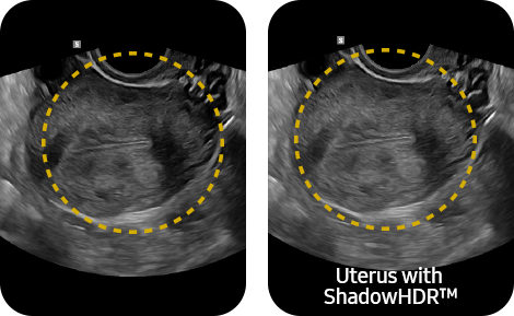
Enhance hidden structures in shadowed regions
ShadowHDR™ selectively applies high-frequency and low-frequency of ultrasound to identify shadow areas where attenuation occurs.
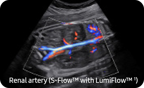
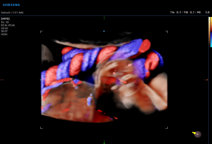
Show blood flow in vessels in a 3D-like display
LumiFlow™ is a function that visualizes Blood flow in 3 dimensional-like to help Understand the structure of blood flow and small vessels intuitively.
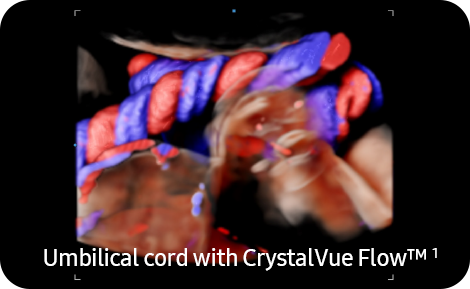
Visualize internal and external structures, and blood flow morphology using volume rendering technologies
CrystalVue™ is an advanced volume rendering technology that enhances visualization of both internal and external structures in a single rendered image.
Reach new diagnostic confidence
with comprehensive tools
Enhance your daily ultrasound diagnosis with the V6, a versatile solution created to efficiently support your clinical demands in general imaging. Benefit from our latest automation tools, which enable you to work with greater ease and achieve reliable results. Our aim is to assist you in prioritizing patient care,
and the V6 stands as an excellent choice.

![]()
An automated classification and annotation of the images
ViewAssist™ a feature based on Deep Learning technology, provides automatic classification of the ultrasound images and annotation of the structures to help healthcare professionals in convenient measurement.
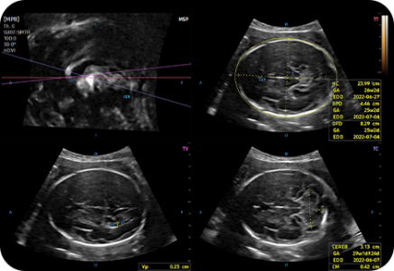
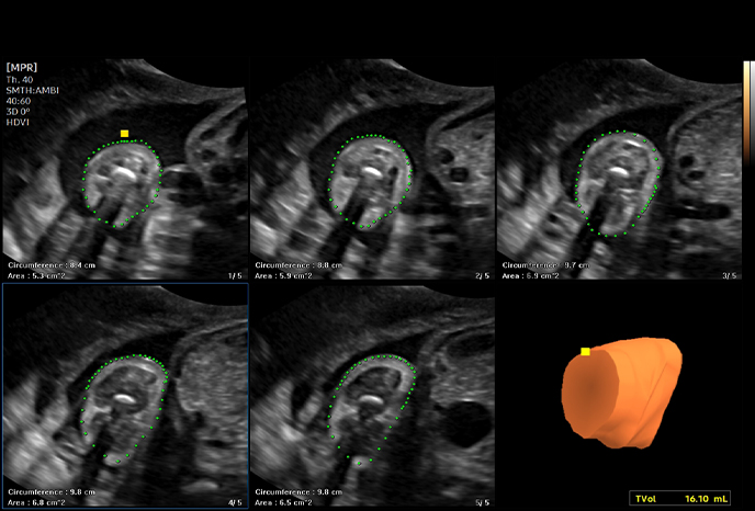
An automated fetal biometry measurement
BiometryAssist™, a feature based on Deep Learning technology, is an automatic technology for biometric measurement. It enables users to measure the fetal growth parameters with one click while maintaining exam consistenc
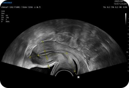
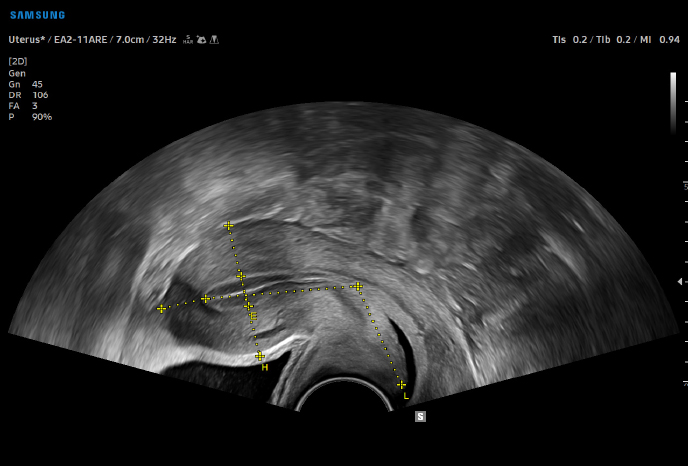
Measure the size and shape of the uterus with AI technology
UterineAssist™ based on Deep Learning technology, automatically measures the size and shape of the uterus, assisting in detecting signs of uterine-related abnormalities, as well as reducing scan time.
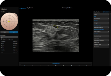
Measure the size of follicles based on 2D imaging
2D Follicle™ ¹ identifies and measures the size of follicles based on a 2D image and provides information about the status during gynecology examinations.
Assess the risk of infertility using volume data
5D Follicle™ identifies and measures multiple ovarian follicles in one scan for rapid assessment of follicular size and status during controlled ovarian stimulation.
Support in deciding delivery method
LaborAssist™ provides information about the progress of delivery from the automatic measurement of the AoP (Angle of Progress) and the direction of the fetal head. This helps in making delivery decisions and effective communication with the mother about the delivery process.
* AoP complies with the metrics specified in the ISUOG Guideline.
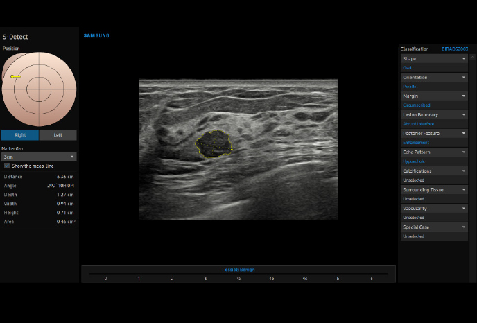
Analyze selected breast lesions and report breast assessment
S-Detect™ for Breast analyzes selected lesions in the breast ultrasound study and shows the analysis data, applies BI-RADS ATLAS* to provide standardized reporting; and helps diagnosis with the streamlined workflow.
* Breast Imaging-Reporting and Data System, Atlas It is a registered trademark of ACR and all rights reserved by ACR.
Analyze selected thyroid lesionsand report thyroid assessment
S-Detect™ ¹,³ for Thyroid analyzes selected lesions in the thyroid ultrasound study and shows the analysis data, provides standardized reporting based on the ATA, BTA, EU-TIRADS, K-TIRADS, and ACR-TIRADS* guidelines; and helps diagnosis with the streamlined workflow.
- ATA: American Thyroid Association
- BTA: British Thyroid Association
- EU-TIRADS: European Thyroid Imaging Reporting and Dat a System
- K-TIRADS: Korean Thyroid Imaging Reporting and Data System
- ACR-TIRADS: American College of Radiology Thyroid Imaging Reporting and Data System
Examine patency of the fallopian tube and morphology of uterus and endometrium
CEUS+ HyCoSy can be used in 3D/4D for effective examination for patency of the fallopian tube and morphology of uterus and endometrium. 4D Prospective storage allows 4D data to be stored at the same time the contrast agent is injected.
Examine fetal heart including blood flow dynamics
5D Heart Color™ identifies 9 standard planes of the heart using fetal STIC data and important information about fetal heart development, complying with AIUM guidelines. It also offers dedicated Preset, Predictive Cursor, Diagnostic Alert, and heart Diastole/Systole timepoints.
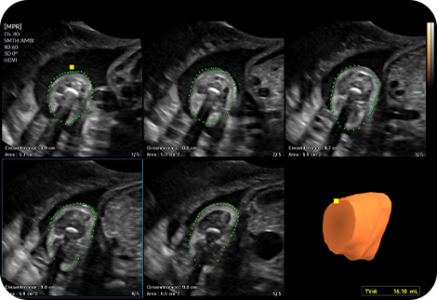
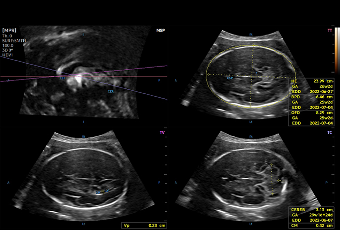
Measure fetal brain with one click
5D CNS+™ ¹ uses intelligent navigation to provide 6 measurements from 3 transverse views of the fetal brain to enhance measurement reproducibility and streamlined workflow.
Estimate fetal weight to check the growth of the fetus
5D Limb Vol.™ is a semi-automated tool to quickly and accurately measure upper arm or thigh volumes from 3 simple seed points on a single volume data set. These measurements can then be used to calculate an accurate estimation of fetal weight.
Display tissue stiffness in color image
A diagnostic ultrasound technique for imaging elasticity, ElastoScan+™ observes the transformation of the tissue strain by the internal or external forces, and converts relative stiffness into a color image.
Easy calculation of the strain ratio between two ROIs
E-Strain™ is designed to enable quick and easy calculation of the strain ratio between two regions of interest for day-to-day practice. Simply by setting the two targets, you can receive accurate, consistent results and make informed decisions in many types of diagnostic procedures.
Classify ovarian tumors
IOTA-ADNEX ¹ is an ovarian tumor classification solution of IOTA Group. Applying the ADNEX model to the system, it can perform all procedures from the initial scan to the final report in the ultrasound diagnosis system.
![]()


 |
 |
 |
 |
|
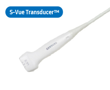
CA1-7ADApplication:Abdomen, Obstetrics, Gynecology, Musculoskeletal, Pediatric, Vascular, Urology |
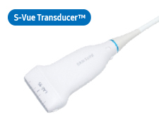
CA3-10AApplication:Abdomen, Obstetrics, Gynecology, Musculoskeletal, Pediatric, Vascular, Urology |
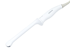
CA1-7SDApplication:Abdomen, Obstetrics, Gynecology |
CA4-10M*Application:Abdomen, Pediatric, Vascular |
 |
 |
 |
 |
|
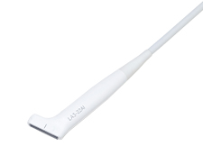
LA2-9S*Application:Small parts, Vascular, Pediatric, Musculoskeletal, Abdomen |
LA3-14ADApplication:Abdomen, Pediatric, Small parts, Vascular, Musculoskeletal |
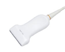
L3-22Application:Musculoskeletal, Pediatric, Vascular, Small parts |
LA3-22AIApplication:Musculoskeletal, Intraoperative |
 |
 |
|||
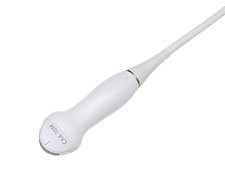
CV1-8AEApplication:Abdomen, Obstetrics, Gynecology |
EV2-10A*Application:Obstetrics, Gynecology, Urology |
 |
 |
 |
||
EA2-11ARE*Application:Obstetrics, Gynecology, Urology |
EA2-11AVE*Application:Obstetrics, Gynecology, Urology |
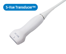
miniER7*Application:Obstetrics, Gynecology, Urology |
 |
 |
 |
||
PA1-5APEApplication:Cardiac, Vascular Abdomen, Pediatric, TCD, Thoracic |
PA4-12BApplication:Cardiac, Pediatric Abdoment, Vascular, TCD |
PA3-8BApplication:Cardiac, Pediatric, Abdomen, Vascular, TCD |
 |
 |
|||
CW6.0Application:Cardiac, Vascular, TCD |
DP2BApplication:Cardiac, Vascular, TCD |
 |
||||
MMPT3-7Application:Cardiac |
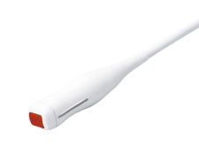
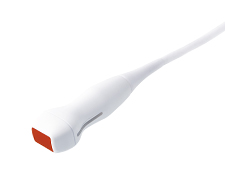
* Ergonomic Transducer (CA1-7S, EA2-11AR, EA2-11AV)
The new convex transducer design with a smooth and slim grip helps users to scan easily and comfortably.
The new endocavity transducer supports natural grip by moving the max width point to a more forward positionand also increased the length of the grip to allow balanced weight distribution.
![]()
![]()