- Description
- Specifications
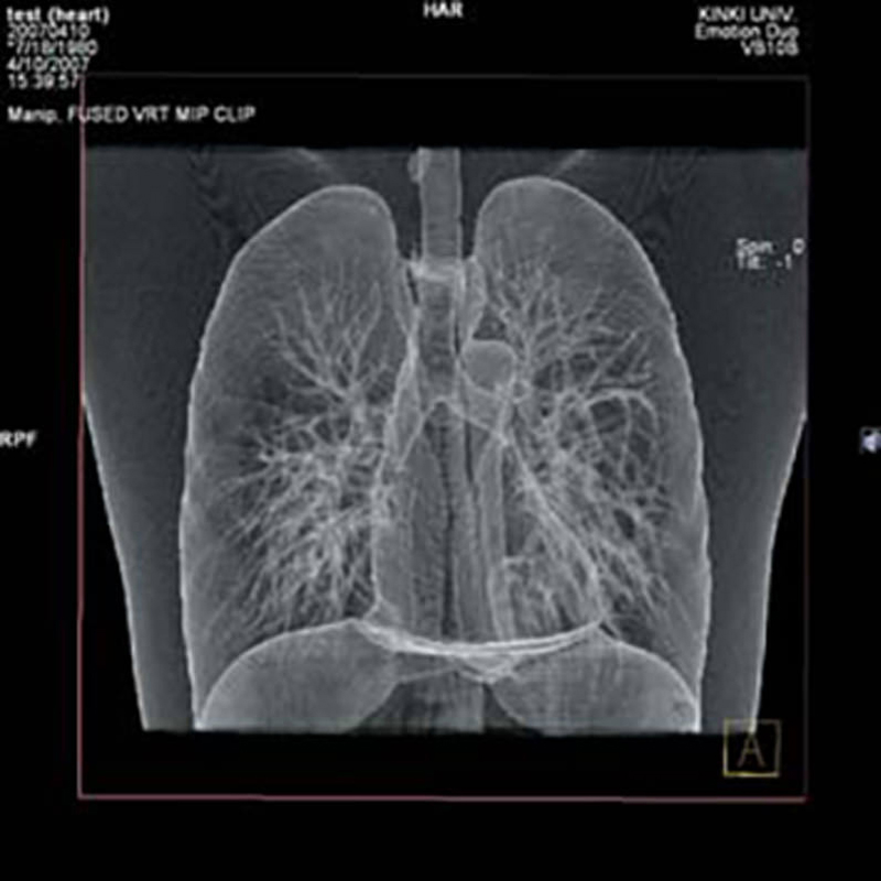
This multipurpose training model can be used for training in x-ray and Computer tomography. It is suitable for training of X-Ray imaging as well as for image interpretation training. Additionally it can be used for assessment of x-ray and CT systems.
Applicable for both plain radiography and CT scanning.
Wide variety of uses in interpretation training, anatomical education, evaluation and assessment of devices and other research.
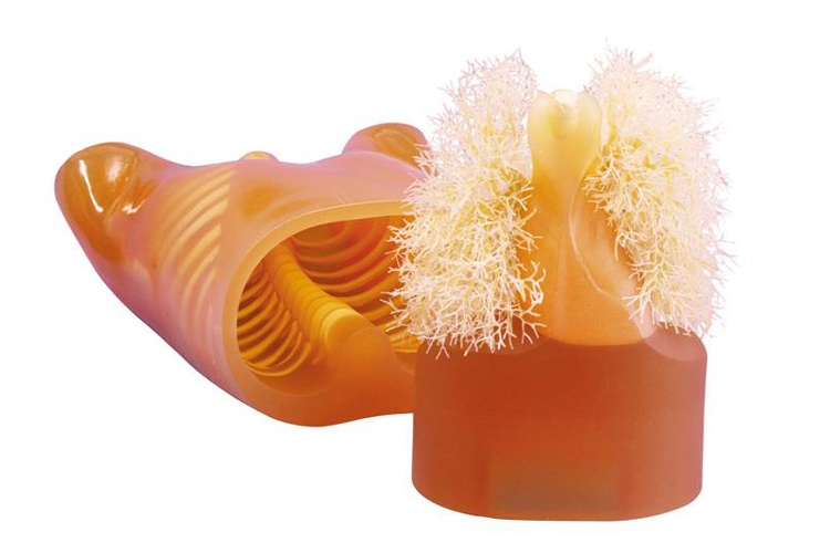
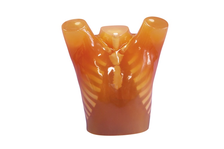
The phantom is an accurate life-size anatomical model of a human torso. The thickness of the chest wall is based on measurement of clinical data. The soft tissue substitute material and synthetic bones have x-ray absorption rates very close to those of human tissues.
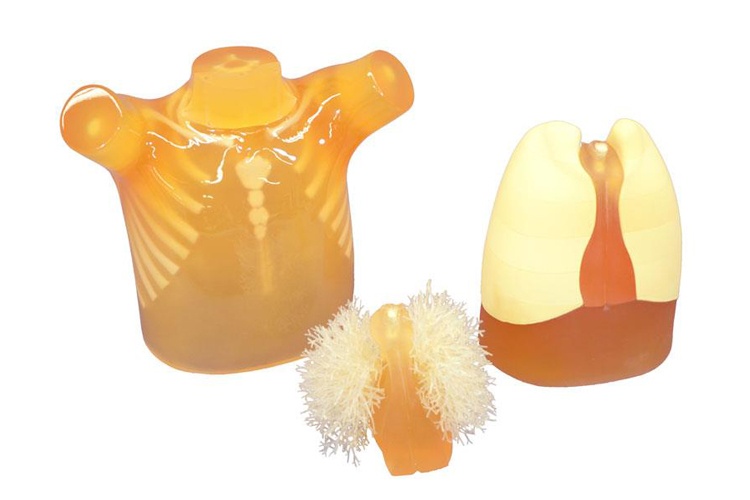
All model structures are made of materials that have x-ray absorption rates close to human tissue. The model can be opened and artificial tumors can be inserted into the lung. 15 different tumors are supplied with the model.
Components for Radioisotope
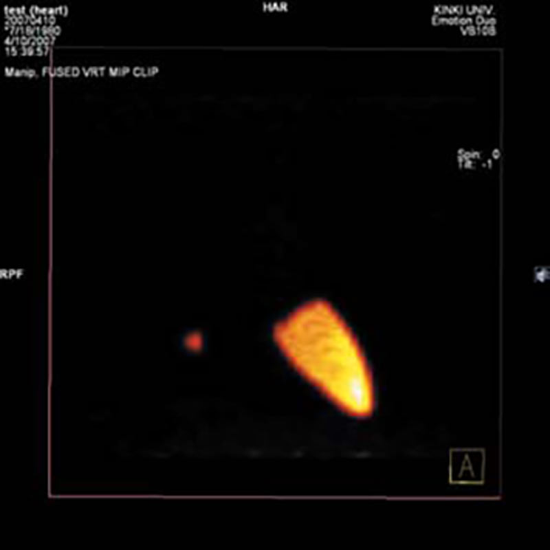
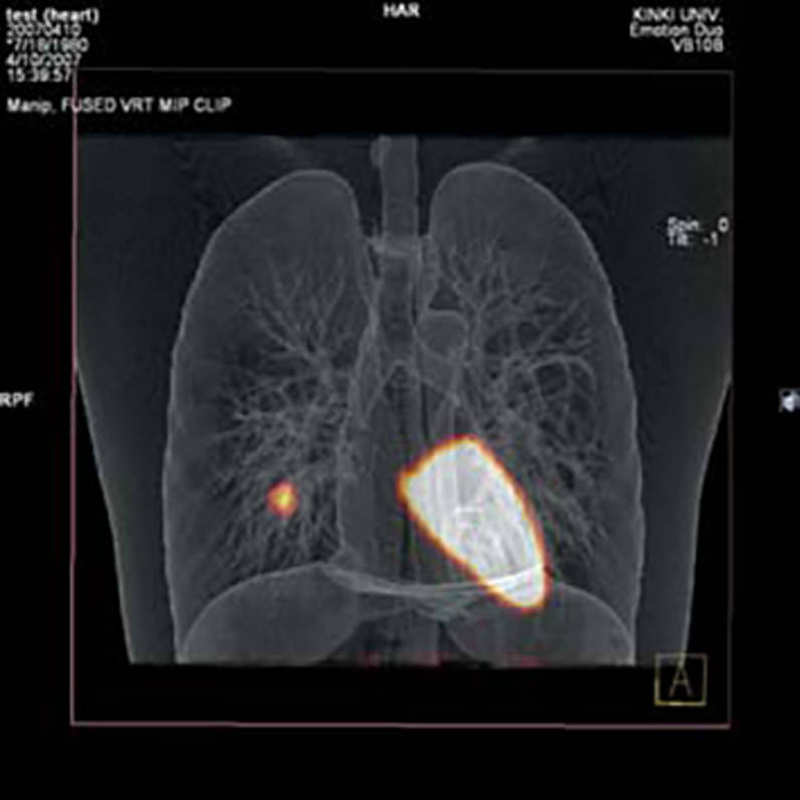
Image fusion experiment with CT & RI can be performed.
The set of RI container inserts can be set in the chest phantom in place of standard inserts allowing wider research applications including PET/CT fusion evaluation. The lungs of urethane foam can be worked easily to accommodate simulated nodules or other inserts.
- Lungs of urethane
- Liver RI container
- Gallbladder RI container
- Pulmonary nodule RI container
- Mediastinum with left myocardium RI container
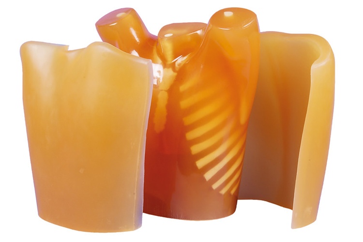
Chest Plate
Chest plates to simulate a larger body type and X-ray absorption. Differences in X-ray absorption depending on body volume can be observed. Using 60mm thick plates, 30mm is added to the front and to the back of the N1 chest.

